|
Getting your Trinity Audio player ready...
|
Introduction
Worth’s four-dot test was first described 100 years ago.
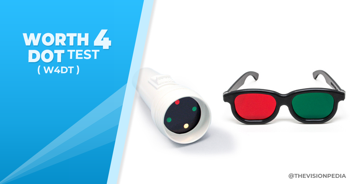
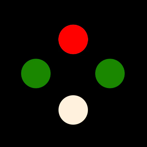
The Worth four dot test is composed of four lights arranged in a diamond shape with a red light at the top, two green lights at the left and right sides, and a white light at the bottom.

Worth four-dot is a gross test and checks for suppression by asking the patient to report the number and color of dots they can see when looking through red-green goggles at four lights or dots of different colors.
The transmission characteristics of the red-green goggles ( Armstrong goggles ) are such that the eye wearing the red filter (usually the right eye) blocks green light to see only the top and bottom light, and the eye viewing through the green filter (usually the left eye) blocks red light and can see the two green lights and the bottom light.
The bottom light is seen in both eyes. A patient with normal binocular vision will appreciate four lights with a flickering red-green light at the bottom because of binocular rivalry.
Therefore, when a patient sees all four dots, one is suspected of having either a normal binocular fusional response with no manifest strabismus or a harmonious anomalous retinal correspondence.
Two Methods of performing worth four dot test
- Distance worth four dot test: target is fixed at a distance of 6 meters from the patient, which may be projected on an illuminated box or the screen.
- Near worth four dot test: target is fixed at a distance of 33cm from the patient, which consists of a flashlight that helps to alter the projection angle.
Requirements to perform worth four dot test
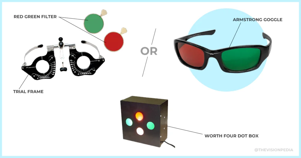
- Trial frame with a red and green filter or red and green filter goggles ( Armstrong Goggle ).
- Illuminated worth four dot box or flashlight.
Assessment of suppression
Normal motor system functioning is essential for binocular vision, but it does not assure that binocular vision exists.
Suppression tests determine whether the patient has the ability to combine the images from both the left and right eye.
If retinal images differ in size due to aniseikonia or in clarity as in amblyopia, anisometropia that is not corrected, or unilateral eye diseases, it could be that the images of the two eyes are not fused because one eye is suppressed.
An inability to appreciate diplopia in some motor system assessments, such as the near point of convergence, may already have suggested suppression.
Simple suppression assessments are also available on the Mallett unit and with some stereopsis tests.
Binocular refraction techniques, such as the Turville infinity balance, can also assess whether gross suppression exists.
Indication of worth four dot test
Manifest strabismus: Helps to identify vertical and horizontal manifest strabismus.
- Esotropia
- Exotropia
- Hypertropia
- Hypotropia
Diplopia: This test can help in determining the nature and the type of diplopia.
- Crossed diplopia
- Uncrossed diplopia
Suppression: It helps to identify which eye is suppressed.
Procedure
- Explain the test and procedure to the patient: “This test tests whether you are using both eyes at the same time to view.
- The patient should wear green-red goggles (Armstrong glasses). The eye with the red filter on its front (usually the left eye) will see the red light, and the eye with the green filter in the front (usually that of the left) will be able to see the green light. Make sure that the patient is not able to view the torch prior to placing the red and green glasses on.
- To test at 6 meters: Ensure that the patient wears their prescribed glasses or contact lenses.
- To test at 33 meters: Hold the Worth 4-dot torch or flashlight in the reading position of the patient in a way that the patient is looking at it from a slight downward angle. For presbyopic patients, make sure the patient is wearing the appropriate refractive correction for near-distance tests. The torch is typically carried by a red light at the top and a white light at the bottom.
- Turn on the Worth 4-dot instrument by keeping the room lights on.
Questions to ask the patient
1) How many lights are you seeing?
2) What color are they?
3) Where are they located?
4) Are all lights in line or some higher than the higher?
5) Do all the lights show up at once, or are they flashing on and off?
There are four possible responses:
- ‘4 dots seen’: Indicates that the patient has normal flat-fusion. This can be verified by asking the question, “How many red dots do I see?” How many are green? Patients will typically see one red, two green, and one yellow dot. The retinal rivalry may cause the white dot to appear yellow or alternate between green and red.
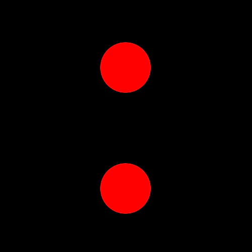
- ‘2 dots seen’: These will be red and white, seen by the patient as two red dots. This indicates suppression of the eye with the green filter in front of it (usually the left). To detect alternating and/or intermittent suppression ask: ‘Are the number of dots changing as you look at them?’ If the number of dots seen is constant, check to see if fusion can be achieved by briefly occluding the nonsuppressed eye.
- ‘3 dots seen’: These will be the two green dots and one white dot, seen by the patient as three green dots. This indicates suppression of the eye with the red filter in front of it (usually the right). To detect alternating and/or intermittent suppression ask: ‘Are the number of dots changing as you look at them?’ If the number of dots seen is constant, check to see if fusion can be achieved by briefly occluding the nonsuppressed eye
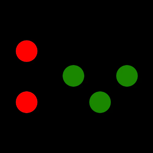
- ‘5 dots seen’: This indicates diplopia. The right eye (usually with the red filter) will see two red dots. The left eye (with the green filter) will see three green dots. Ask the patient to indicate where the red dots are in relation to the green ones. If the red dots (usually seen by the right eye) are to the right of the green dots, this indicates uncrossed diplopia and an eso deviation. If the red dots are to the left of the green dots, this indicates crossed diplopia and an Exo deviation. If the red dots are below the green dots, this indicates an R/L deviation. If the red dots are above the green dots, this indicates an L/R deviation.
- Repeat the testing with room lights off if suppression or diplopia is found.
- If there is suppression at a distance but not near, measure the extent of the suppression scotoma by moving the near target away from the patient and asking them to report when suppression occurs.
- In patients who show suppression, it can be useful to repeat the test with the red-green goggles reversed to ensure an accurate assessment (Simons & Elhatton 1994).
Note: Children who are unable to communicate verbally can be asked to touch dots to indicate the number of dots. Touching four indicates normal flat-fusion. Some evidence suggests that the test can detect suppression, but it will not distinguish between normal fusion or alternating suppression. (Lueder and Arnoldi 1996)
Worth Four Dot Test Interpretation
4 dots- 1 Red, 2 Green, 1 Orange
Final interpretation: Binocular vision
3 Green dots
Final interpretation: Right eye Suppression.

2 Red dots
Final interpretation: Left eye Suppression.
3 Green dots & 2 Red dots Alternatively
Final interpretation: Alternate Suppression.

5 dots, 2 Red in the left eye, 3 Green in the right eye
Final interpretation: Uncrossed Diplopia.

5 dots, 3 Green dots in the left eye, 2 Red dots in the right eye
Final interpretation: Crossed Diplopia.
5 dots, 2 Red on the upper side and 3 Green on the lower side
Final interpretation: Vertical Diplopia with Right Hypotropia or Left Hypertropia.

5 dots, 3 Green on the upper side and two Red on the lower side
Final interpretation: Vertical Diplopia with Left Hypotropia or Right Hypertropia.

Most common errors
- Conducting the test using the patient’s eyes unaided or when they are suffering from poor visual acuity due to their glasses.
- Assuming that the absence of suppression confirms the presence of stereopsis
Advantages
- The Worth four-dot test is widely accessible, inexpensive, affordable, and easy to utilize. It is a great test to test fusion at a distance or near.
- It provides an indication of suppression in the sense that other tests may reveal the presence of suppression when the 4-dot test suggests that none is present. This is particularly true for near 4-dot testing because of the relatively large angular size of the lights when viewed near compared to distance viewing.
- Conversely, the rivalry produced by the red-green Assessment of Binocular Vision 197 goggles may lead to dissociation even in a patient with good or normal binocular vision so the test can suggest the existence of suppression when none is present under habitual viewing conditions.
- It is relatively easy to record and understand the results.
- It can be quick and straightforward to perform in the clinic as the test is easy to orientate, and the red-green goggles are put over the eyes.
- It is not as dissociative as the cover test.
- It differentiates monocular diplopia from binocular diplopia.
- It can also be employed with prisms before surgery to assess the risk of postoperative diplopia and provide indications as to whether there are torsion or visual field defects.
Disadvantages
- Subjective in nature and depends on the responses of patients.
- The color blind can’t accurately take the test because the colors used in the test are green and red.
- If performing the test twice, for example, near and at a distance, the patient (especially children) may remember their previous answer and give the same answer from the last test, providing inaccurate results.
- The patient needs to have fusion and stereopsis in order to obtain exact results.
- The main drawback of testing is the brightness of the green and red targets may vary significantly in different tests, and so can how the goggles transmit green and red light, which means that the result of the test can differ according to whether the goggles are worn in the traditional format (red goggles placed on one’s right eye) and reversed (Simons and Elhatton, 1994).
- Another drawback of this test is a person with constant strabismus, as well as an irregular retinal correspondence, could get an acceptable result. Positive test results cannot necessarily guarantee the existence of normal binocular vision.
- It is an invaluable test when used to evaluate long-standing and acquired strabismus in adults and manage complex diplopia.
References
- David. B. Elliott. Clinical Procedures in Primary Eye Care 3rd Edition – SILO.PUB. https://silo.pub/clinical-procedures-in-primary-eye-care-3rd-edition.html
- Eunoo Bak, Hee Kyung Yang, Jeong-Min Hwang. Validity of the Worth 4 Dot Test in Patients with Red-Green Color Vision Defect, Optom Vis Sci. 2017 May;94(5):626-629
- https://en.wikipedia.org/wiki/Worth_4_dot_test
- https://eyewiki.aao.org/Worth_4_dot

